If you’ve been diagnosed with rectal cancer, further testing is necessary to determine the extent of the tumor. In experienced hands, a rectal MRI is one of the most accurate tools for local staging. It also plays a pivotal role in pre-surgical assessment and treatment planning.
To understand the nuances of rectal cancer diagnosis and staging, we spoke with MRI expert, Dr. Vikram Hatti. From detection to post-treatment, Dr. Hatti shares the benefits of rectal MRI and offers advice for patients who are navigating a rectal cancer diagnosis.
DocPanel is committed to making sure every patient receives excellent care. If you would like an expert second opinion on your medical imaging scan from Dr. Hatti or one of our other subspecialty radiologists, you can learn more here.
Rectal Cancer Detection
[DocPanel] What are the first steps in determining whether a rectal tumor is benign (non-cancerous) or malignant (cancerous)?
[Dr. Hatti]
A colonoscopy is usually the first test performed after a rectal polyp or tumor has been detected. During the colonoscopy, a tissue sample of the growth is collected for laboratory analysis to determine whether or not it is cancerous. (There are some instances where the placement of the polyp does not allow for biopsy, but most of the time it is possible.) A PET-CT scan may also be ordered as an alternate (or additional) way to assess the nature of a rectal tumor.
Rectal polyps can be divided into three categories:
- benign, noncancerous rectal polyps
- precancerous rectal polyps (i.e. noncancerous tumors that have the – potential to convert into rectal cancer)
- malignant, cancerous rectal tumors
Rectal Cancer Staging
[DocPanel] What role does rectal MRI play in the staging process?
[Dr. Hatti]
Typically, a rectal MRI scan is performed after it has already been proven by biopsy that the tumor is cancerous. It’s the most helpful imaging tool available to diagnose rectal cancer because of its ability to capture detailed pictures of the tissue layers within the rectal wall. This allows radiologists to determine the extent of the rectal tumor, allowing for accurate rectal cancer staging. A rectal MRI scan also shows the lymph nodes near the rectum, which aids in assessing the possibility of lymph node involvement.
PET-CT and transrectal sonogram may also be helpful in certain cases.
[DocPanel] How is the stage of rectal cancer determined?
[Dr. Hatti]
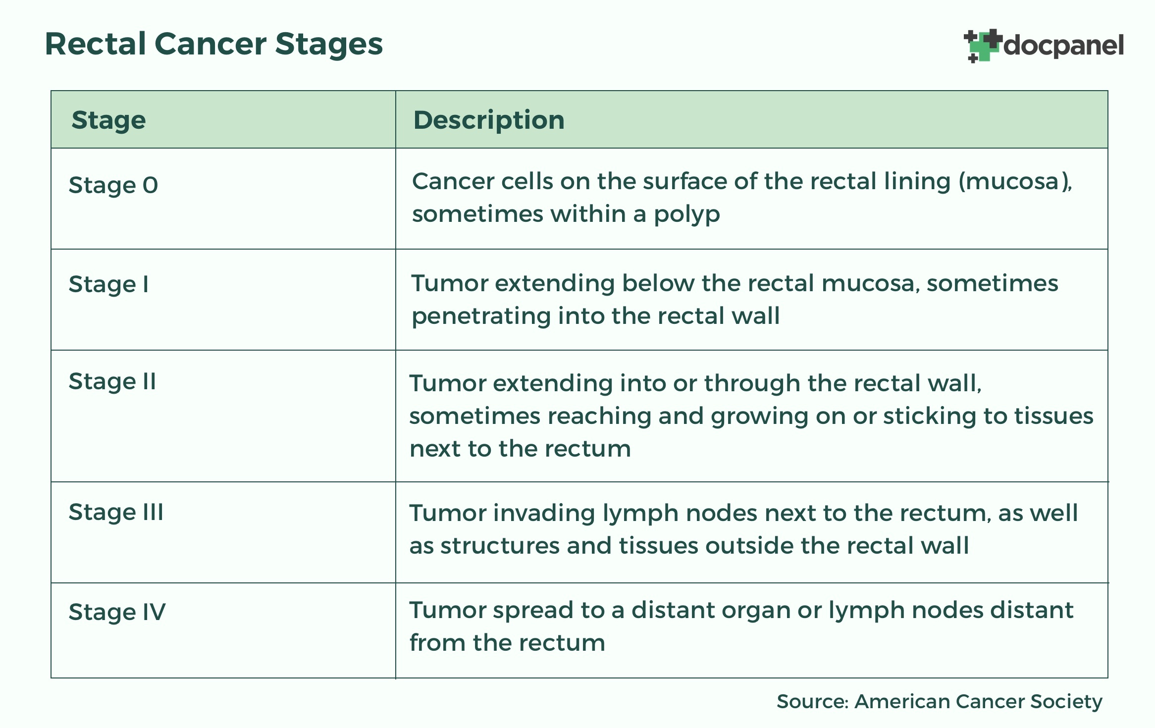
Rectal cancers fall into one of five possible stages (stage 0 through stage 4). The lower the number, the less the cancer has spread.

Rectal cancer is staged using the TNM staging criteria. This includes taking the following into consideration:
- T – how far the cancer has grown/infiltrated into the wall layers of the rectum
- N – whether or not it has spread to nearby lymph nodes
- M – the spread (metastasis) to distant lymph nodes or other organs
Numbers and letters are assigned to the T, N, and M categories to provide details about each factor. This information is then combined in a process called stage grouping to assign an overall stage. It’s a complex process, so it’s important to have your doctor explain your stage in full detail.
Rectal Cancer Treatment
[DocPanel] What role does rectal MRI play in treatment planning?
[Dr. Hatti]
As with staging, a rectal MRI is the best imaging test for determining which type of treatment can and should be used. The major treatments are total mesorectal excision (TME), neoadjuvant radiotherapy (radiation), and chemotherapy. Oftentimes, more than one treatment type is required to treat rectal cancer. Whether a patient with rectal cancer is a candidate for total mesorectal excision (TME) only or neoadjuvant therapy followed by TME, is made based on the findings on MRI.
Patients with T1, T2, and T3 N0 lesions less than 5 mm invasion can undergo total mesorectal excision. Patients with T3 N1 involvement greater than 5 mm invasion can undergo TME as short term radiation therapy. Patients with T3 that are less than 1 mm to the mesorectal fascia and T4 N2 would undergo neoadjuvant chemotherapy and long-term radiation therapy.
[DocPanel] What role does MRI play in post-treatment for rectal cancer survivors?
[Dr. Hatti]
Post-treatment, rectal MRI scans help screen and assess for the presence of subtle areas of recurrence of the tissue surrounding the rectum and nearby lymph nodes. These should be performed regularly, to ensure any new cancer growth is caught early.
Advice for Navigating a Rectal Cancer Diagnosis
[DocPanel] What advice do you have for patients navigating a rectal cancer diagnosis?
[Dr. Hatti]
There are challenges in diagnosing rectal cancer. For instance, it is very difficult to differentiate between the mesorectal fascia, the rectal wall, and the luminal portion of the rectum. Determining whether or not there is an infiltration of the fascia or adjacent pelvic organs is also very complicated. So staging is not an easy or simple process. It requires great skill from the radiologist.
With this in mind, my advice would be as follows:
Consult with an oncologist who has vast experience treating rectal cancer.
Get a second opinion on your rectal imaging scans
Post-treatment, continue to get periodic screening MRI scans of the rectum to assess for the presence of recurrence.
