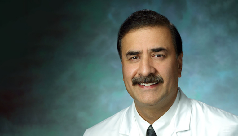
Academic Neuroradiologist, Spine Interventionist, and Radiologist at DocPanel, Dr. Majid Khan, has earned worldwide recognition for his expertise and deep dedication to the field.
A leading specialist in spine tumor ablation, Dr. Khan has performed the most cases of osseous microwave tumor ablation in the world. He’s also an expert in spine cement augmentation procedures, like kyphoplasty and vertebroplasty for treatment of compression fractures, and is an integral member of the Jefferson Headache Center which is a national referral center for treatment of spontaneous CSF leak and Tarlov cyst patients.
In this exclusive interview with Dr. Khan, we dive deeper into his expertise. From common pitfalls in diagnosing tumors in the head, neck, and spine to advice on how to overcome such challenges, Dr. Khan gives us a glimpse into his work with exciting reveals about his latest projects.
[DocPanel] You’ve performed the highest number of spine tumor ablations in the US – that’s an incredible accomplishment. As an expert in this area, what would you say are some of the major challenges involved with this procedure? What advice can you offer to overcome them?
[Dr. Khan] Throughout my career I have worked in University Hospitals and Academic Centers, so I’ve always been well-placed to do these types of interventions – knowing that I have good neurosurgical cover if needed.
In my opinion, the most important part of oncological treatment planning is having a strong multidisciplinary team. All the relevant players need to be sitting at the table to discuss the patient’s clinical picture, and decide on the best treatment modality and follow-up plan. When you are ablating tumors close to the spinal cord and/or any nerves, you need to be very confident in the machine that you are using for ablation because minimal error can have devastating effects. Protective mechanisms used during the case have to be known well before starting such procedures. There is definitely a learning curve; training with someone who does these procedures regularly is a big plus, especially during a fellowship.
I would advise people who want to start spine tumor ablations that they should get good training and also perform a few cases in cadaveric labs to get used to the machine and its workings.
[DocPanel] What are some of the pitfalls in diagnosing tumors in the head, neck, and spine? In your experience, do you see many patients who were initially misdiagnosed?
[Dr. Khan] Head and neck imaging is very challenging. A good training program in which the physician trainee gets to see a number of these cases is very helpful. There is a saying in radiology that, "what mind doesn’t know, eyes don’t see" – if you are not exposed to some of these rare entities during your training, it becomes hard to appreciate them in clinical practice.
Having an internal checklist is very important as you are going through complicated scans in this region of the body. Comparisons, if available, should always be looked at. Good practice in oncological imaging is to compare at least 2-3 prior scans with the present scan, if available, rather than just comparing the most immediate prior scan. A small but definite change can have big treatment implications.
Unfortunately, missed diagnoses are seen across almost every specialty of radiology. Fortunately, most of these misses can be caught with repeat scans or when another radiologist interprets the images. They may also come to light early on by peer review metrics kept in place by most practices.
[DocPanel] What are some of the latest developments within advanced head, neck, and spine imaging that you’re especially excited about?
[Dr. Khan] The field of radiology is changing every day with modalities becoming available for better diagnoses and treatment. In the field of neuroradiology, diffusion and perfusion imaging of head and neck tumors has been a game-changer in regard to differentiating aggressive versus nonaggressive tumors. This is further aided by the availability of MR spectroscopy.
Functional MRI imaging of the brain is very precise and extremely helpful to neurosurgeons. It helps guide the surgery in real-time based on the functional data obtained beforehand. CSF flow studies are very helpful in certain situations, as are the different advanced myelographic techniques such as dynamic spine myelograms and dynamic subtraction myelograms which are used to diagnose subtle CSF leaks in patients with intractable headaches. Diffusion tensor imaging is another example of an advanced imaging technique used to evaluate both the brain and spinal cord. There are also multiple new techniques on the horizon for patients with epilepsy and demyelinating disease processes.
[DocPanel] Are you currently working on any projects that you can share a preview of with us?
[Dr. Khan] There are a few ongoing projects at the moment, one of which is long-term local regional control of the spinal osseous metastasis after tumor ablation with almost a two-year follow-up done on such patients.
I have also worked on several recent publications. One compares an MRI technique used to diagnose osteoporosis with both DEXA scan and CT densitometry, showing a very good correlation. We also recently published a paper demonstrating US academic center experience with using implant kyphoplasty (spine Jack) in patients with spinal compression fractures. Another recently published paper I worked on covers rare non-squamous cell tumors of the head and neck region, showcasing 20 years of experience with such tumors from a single institute.
[DocPanel] What inspired you to get into the field of radiology?
[Dr. Khan] I chose to get into the field of radiology because it combines the best of both worlds when it comes to patient care. I get to spend time with colleagues, having stimulating conversations where we discuss, for example, the imaging aspects of a complicated case and come up with differential diagnoses. And, on the other hand, I’m also able to practice interventional radiology, which allows me to incorporate that all-important patient contact and healing touch.
[DocPanel] At what point did you feel a pull toward neuroradiology?
[Dr. Khan] I became very intrigued by brain and spine anatomy early on in my
radiology residency. During my first year, I developed such a strong pull toward neuroradiology over all other specialties – I was certain I would pursue a fellowship in it. There is so much that we don’t know about the brain, and, every day, we continue to learn new things about the neuro-spinal axis. It’s fascinating.
Dr. Majid Khan is an Interventional Neuroradiologist at Johns Hopkins, an Associate Professor of Radiology at Thomas Jefferson University Hospitals, and Radiologist at DocPanel. He is also Director of Non-Vascular Spine Intervention at Johns Hopkins University hospitals. Dr. Khan graduated from the University of Kashmir, India in 1993. He completed his residency in radiology at the Nassau University Medical Center at Stony Brooks University and then completed his fellowship in Neuroradiology at Johns Hopkins University. Dr. Majid enjoys teaching and is especially interested in advanced head & neck and spine tumor imaging, with extensive experience in performing spine tumor ablations and other spinal interventions.
