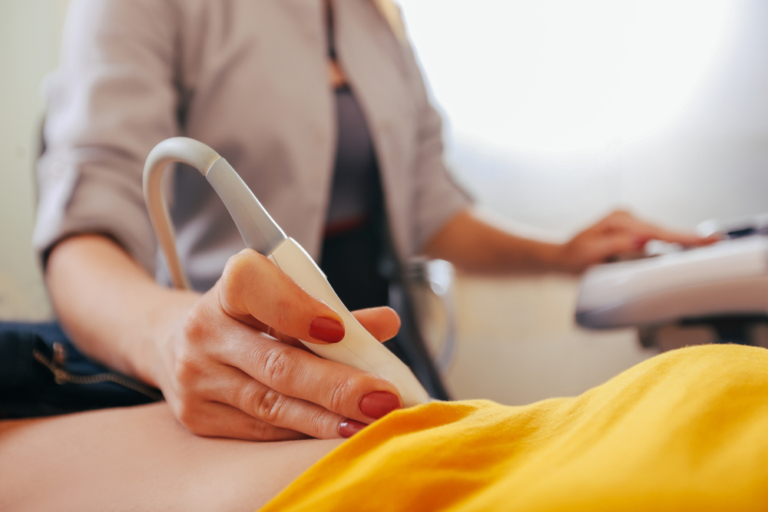
Ultrasound is an excellent front-line diagnostic tool for evaluating the liver.
It can help assess the presence of liver disease (such as fatty liver), detect liver lesions, and much more. But many factors can affect the accuracy of your diagnosis.
We spoke with Dr. Barbara McComb, a Mayo Clinic diagnostic radiologist and ultrasound expert with over 38 years of experience, to find out what patients should be aware of when getting a liver ultrasound. Here, she explains how getting a second opinion can help provide clarity on your diagnosis, followed by a Q&A with the most common questions patients ask about their liver ultrasound.
Benefits of a Liver Ultrasound Second Opinion
When you get a liver ultrasound, your radiology report will be used by your doctors to make important decisions about your diagnosis and how to proceed. Having a subspecialist provide a second opinion can help confirm that findings reported have been accurately assessed. It may also help you and your doctors determine whether follow-up screening or treatment is needed. If you have questions about your results or are feeling worried about an abnormal finding, a second opinion is also a great way to get more clarity on your diagnosis and peace of mind moving forward.
3 Areas a Liver Ultrasound Second Opinion Can Help Ensure Accuracy
1. Checking image quality
A liver ultrasound study typically contains a series of multiple images taken by the technologist performing the examination. Because this type of imaging test is done free-hand, it is very user-dependent. The areas imaged and quality of imaged taken depend greatly on visibility and technique. Compromised images can negatively impact what can be seen by the radiologist. A second opinion can help confirm that the images are of high enough quality to make an accurate diagnosis.
2. Diagnosing and characterizing liver disease
Ultrasound is useful for distinguishing liver cysts from solid masses. Some masses have unique ultrasound features while others share similar characteristics on ultrasound. A liver ultrasound is also used to assess for diffuse liver disease, including fatty liver. A radiologist providing a second opinion can help characterize a difficult liver lesion, evaluate the liver for the presence of fat, and determine other tests that might be necessary to assess the liver.
3. Determining whether follow-up imaging is necessary
An ultrasound may be the first screening test performed when evaluating the liver. Depending on the results, your doctor may recommend another examination. For example, a CT, MRI, or biopsy may be indicated to differentiate between liver fibrosis and fat, further quantify the severity of fat, or evaluate for other disease in the liver. CT or MRI may also be recommended to further evaluate certain liver lesions. A radiologist providing a second opinion can help guide you about additional testing.
Understanding Your Liver Ultrasound
What symptoms indicate a liver ultrasound is needed?
[Dr. McComb] A wide variety of signs, symptoms, and laboratory findings can be associated with liver disease and may prompt the ordering of an ultrasound study. Some examples include unexplained weight loss, abdominal discomfort or swelling, early satiety (feeling full without eating much), nausea or vomiting, weakness, jaundice, palpable mass, coagulation problems, and liver or spleen enlargement on a physical examination.
What does a liver ultrasound scan show?
[Dr. McComb] Ultrasound is excellent at showing normal anatomy and the presence of abnormalities in the liver. It is particularly excellent for differentiating cysts from solid masses. Simple cysts have a thin wall and contain fluid, which shows up as a darker center than solid masses have on ultrasound. Complex cysts may have associated lumps (nodules), calcifications, or multiple tissue bands. Solid masses can even be evaluated for blood flow by a technique called Doppler ultrasound, and cystic areas within masses can be distinguished from the solid parts.
Ultrasound can also evaluate diffuse liver diseases, such as fatty liver, hepatitis, and cirrhosis. For example, a fatty liver (steatosis) is typically brighter (more “echogenic” or “hyperechoic”) on a liver ultrasound than normal liver, while hepatitis may be less bright (“hypoechoic”). A cirrhotic liver often looks shrunken and lumpy. A special technique called elastography can be added to an ultrasound study to help measure the elasticity of the liver and assess the severity of fibrosis.
Dilated bile ducts and any fluid near the liver (ascites, fluid collections) will also typically show up on a liver ultrasound. Other organs, including the gallbladder, right kidney, and at least a portion of the pancreas are often seen as well.
Can a liver ultrasound detect liver cancer?
[Dr. McComb] Ultrasound can detect many cancerous (malignant) and non-cancerous (benign) liver masses. A mass is generally more easily seen by ultrasound the more it differs in appearance, and therefore stands out, from background tissue. Although not a hard-fast rule, in many cases a larger mass is more easily detected than a smaller mass.
But the detection of any mass is only possible if there is a good ultrasound “window.” Unfortunately, bone, calcification, and gas act as barriers to a liver ultrasound beam. Fatty tissue in front of or within the liver also reduces the penetration of the beam and the visibility of masses.
Another thing to recognize is that there is some overlap in the appearances of cancerous and non-cancerous masses. Fortunately, contrast-enhanced ultrasound (CEUS) has fairly recently been emerging in the United States as a useful technique to further evaluate liver lesions not well characterized by conventional ultrasound. It has been used in Europe and Asia for several years.
With CEUS, a microbubble contrast agent is injected into a vein during the liver ultrasound study to help better visualize blood flow. Analysis of flow patterns is useful in differentiating certain lesions from one another, as well as in following certain lesions.
The liver is the largest solid organ in the body – does this make it a more difficult area to image?
[Dr. McComb] The size of the liver compared to other organs does not impede liver ultrasound imaging. Liver tissue is very well suited to ultrasound evaluation as long as there is a good “window” for the ultrasound beam and it can penetrate sufficiently. Technologists performing liver ultrasound are experts at working with patients to get the best possible images.
For example, since ultrasound cannot pass through ribs, a technologist may ask a patient to take a deep breath and/or roll to bring more of the liver down below the ribs so that it can be better seen.
What terms on a liver ultrasound report should a patient be aware of?
[Dr. McComb] Many different terms are used in liver ultrasound reports to describe both normal liver and liver abnormalities. Radiologists frequently describe the “echogenicity” and “echotexture” of background liver tissue and report any lesions (such as cysts, solid masses, etc) visible on the examination.
They may describe whether a particular lesion is darker than (hypoechoic), similar to (isoechoic), or brighter than (echogenic) background liver tissue. Margins of the lesion are often described (smooth, irregular, etc) as well as any visible blood flow within the lesion on Doppler ultrasound, if performed. The presence of any abnormal enlargement (dilatation) of bile ducts in or outside the liver will be reported, along with any abnormal fluid (ascites or fluid collection) next to the liver. Other organs or portions of them can also be seen while scanning the liver, and a patient may see gallbladder or kidney stones (calculi) reported.
What does it mean to have an ‘echogenic liver’? What are the possible causes?
[Dr. McComb] A liver is described as “echogenic” by a radiologist when the echoes reflected from the ultrasound beam appear brighter (whiter) than those from a normal liver.
This appearance most commonly indicates the presence of fatty liver (steatosis), although steatosis can be overestimated by attenuation of the ultrasound beam by overlying soft tissue fat. Increased echogenicity can also sometimes be associated with cirrhosis and chronic hepatitis. In these cases, the liver echotexture may also be described as abnormally coarse. Other diseases that infiltrate or deposit in the liver may also increase the echogenicity, including certain storage and infectious diseases.
